The Gallbladder
The Gallbladder
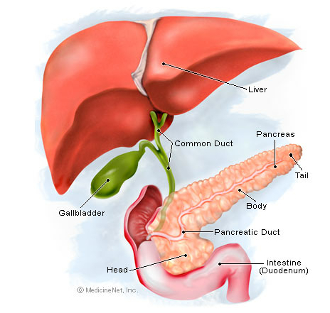
The gallbladder is a small pouch that sits just under the liver. The gallbladder stores bile produced by the liver. After meals, the gallbladder is empty and flat, like a deflated balloon. Before a meal, the gallbladder may be full of bile and about the size of a small pear.
In response to signals, the gallbladder squeezes stored bile into the small intestine through a series of tubes called ducts. Bile helps digest fats, but the gallbladder itself is not essential. Removing the gallbladder in an otherwise healthy individual typically causes no observable problems with health or digestion yet there may be a small risk of diarrhea and fat malabsorption.
Gallbladder Conditions
Gallstones (cholelithiasis): For unclear reasons, substances in bile can crystallize in the gallbladder, forming gallstones. Common and usually harmless, gallstones can sometimes cause pain, nausea, or inflammation.
Cholecystitis: Inflammation of the gallbladder, often due to a gallstone in the gallbladder. Cholecystitis causes severe pain and fever, and can require surgery when inflammation continues or recurs.
Gallbladder cancer: Although rare, cancer can affect the gallbladder. It is difficult to diagnose and usually found at late stages when symptoms appear. Symptoms may resemble those of gallstones.
Gallstone pancreatitis: An impacted gallstone blocks the ducts that drain the pancreas. Inflammation of the pancreas results, a serious condition.
Gallbladder Tests
Abdominal ultrasound: a noninvasive test in which a probe on the skin bounces high-frequency sound waves off structures in the belly. Ultrasound is an excellent test for gallstones and to check the gallbladder wall.
HIDA scan (cholescintigraphy): In this nuclear medicine test, radioactive dye is injected intravenously and is secreted into the bile. Cholecystitis is likely if the scan shows bile doesn’t make it from the liver into the gallbladder.
Endoscopic retrograde cholangiopancreatography (ERCP): Using a flexible tube inserted through the mouth, through the stomach, and into the small intestine, a doctor can see through the tube and inject dye into the bile system ducts. Tiny surgical tools can be used to treat some gallstone conditions during ERCP.
Magnetic resonance cholangiopancreatography (MRCP): An MRI scanner provides high-resolution images of the bile ducts, pancreas, and gallbladder. MRCP images help guide further tests and treatments.
Endoscopic ultrasound: A tiny ultrasound probe on the end of a flexible tube is inserted through the mouth to the intestines. Endoscopic ultrasound can help detect choledocholithiasis and gallstone pancreatitis.
Abdominal X-ray: Although they may be used to look for other problems in the abdomen, X-rays generally cannot diagnose gallbladder disease. However, X-rays may be able to detect gallstones.
Gallbladder Treatments
Gallbladder surgery (cholecystectomy): A surgeon removes the gallbladder, using either laparoscopy (several small cuts) or laparotomy (traditional “open” surgery with a larger incision).
Antibiotics: Infection may be present during cholecystitis. Though antibiotics don’t typically cure cholecystitis, they can prevent an infection from spreading.
Chemotherapy and radiation therapy: After surgery for gallbladder cancer, chemotherapy and radiation may be used to help prevent cancer from returning.
Ursodeoxycholic acid: In people with problems from gallstones who are not good candidates for surgery, this oral medicine is an option. Ursodeoxycholic acid may help dissolve small cholesterol gallstones and reduce symptoms. Another oral solution is called Chenix.
Extracorporeal shock-wave lithotripsy: High-energy shockwaves are projected from a machine through the abdominal wall, breaking up gallstones. Lithotripsy works best if only a few small gallstones are present.
Contact solvent dissolution: A needle is inserted through the skin into the gallbladder, and chemicals are injected that dissolve gallstones. This technique is rarely used.