The Spleen
The Spleen
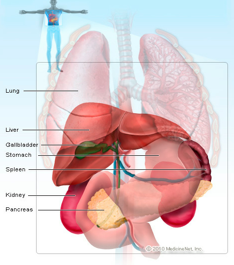
The spleen is an organ in the upper far left part of the abdomen, to the left of the stomach. The spleen varies in size and shape between people, but it’s commonly fist-shaped, purple, and about 4 inches long. Because the spleen is protected by the rib cage, you can’t easily feel it unless it’s abnormally enlarged.
The spleen plays multiple supporting roles in the body. It acts as a filter for blood as part of the immune system. Old red blood cells are recycled in the spleen, and platelets and white blood cells are stored there. The spleen also helps fight certain kinds of bacteria that cause pneumonia and meningitis.
Spleen Conditions
Enlarged Spleen (Splenomegaly): An enlarged spleen, usually caused by viral mononucleosis (“mono”), liver disease, blood cancers (lymphoma and leukemia), or other conditions.
Ruptured spleen: The spleen is vulnerable to injury, and a ruptured spleen can cause serious life-threatening internal bleeding and is a life-threatening emergency. An injured spleen may rupture immediately after an injury, or in some cases, days or weeks after an injury.
Sickle cell disease: In this inherited form of anemia, abnormal red blood cells block the flow of blood through vessels and can lead to organ damage, including damage to the spleen. People with sickle cell disease need immunizations to prevent illnesses their spleen helped fight.
Thrombocytopenia (low platelet count): An enlarged spleen sometimes stores excessive numbers of the body’s platelets. Splenomegaly can result in abnormally few platelets circulating in the bloodstream where they belong.
Accessory spleen: About 10% of people have a small extra spleen. This causes no problems and is considered normal.
Spleen Tests
Physical examination: By pressing on the belly under the left ribcage, a doctor can feel an enlarged spleen. He or she can also look for other signs of illnesses that cause splenomegaly.
Computed tomography (CT scan): A CT scanner takes multiple X-rays, and a computer creates detailed images of the abdomen. Contrast dye may be injected into your veins to improve the images.
Ultrasound: A probe is placed on the belly, and harmless sound waves create images by reflecting off the spleen and other organs. Splenomegaly can be detected by ultrasound.
Magnetic resonance imaging (MRI): Magnetic waves create highly detailed images of the abdomen. By using contrast dye, blood flow to the spleen can also be measured with MRI.
Bone marrow biopsy: A needle is inserted into a large bone (such as the pelvis) and a sample of bone marrow is taken out. Leukemia or lymphoma, which cause splenomegaly, are sometimes diagnosed by bone marrow biopsy.
Liver and spleen scan: A small amount of radioactive dye is injected into the arm. The dye moves throughout the body and is collected in both of these organs.
Spleen Treatments
Splenectomy: The spleen is removed by surgery, either through laparoscopy (multiple small incisions) or laparotomy (one large incision).
Vaccinations: After spleen removal, it’s important to get vaccinations against certain bacteria, such as H. influenza and S. pneumonia. An absent spleen increases vulnerability to these infections.
Usually, treatments for spleen conditions focus not on the spleen, but on treating the underlying condition.